


Physics for the 21st Century
Biophysics Interview with Featured Scientist Vinothan N. Manoharan

Interviewer: What are your specific research interests?
VINOTHAN: My particular research and interest now has to do with systems of many particles that are interacting at once. I’m interested in this very general problem, which is: “How do many particles come together?” and also “How do they come together to form some organized whole?” A comparable question in biology might be: “How do proteins come together to form a virus?” and in condensed matter physics: “How do many atoms come together to form a crystal?” We know a lot about the individual properties of atoms, proteins, and these colloidal particles that I work with. We know a lot about their individual properties; but when you put a bunch of them together, they do interesting things that we don’t expect.
Interviewer: What is a “colloid”?
VINOTHAN: Most of the systems I work with are colloidal systems, which means they consist of some particles floating around, suspended in an immiscible fluid. The particles are larger than the scale of a molecule—so they may be from 10 nanometers on up. The largest size is set by the size that would stay suspended in the fluid for a long time just due to thermal fluctuations or Brownian Motion—10 microns or so.
Interviewer: Can you tell me what self-assembly is and why self-assembly is important?
VINOTHAN: The dream is that some day we’ll be able to throw a bunch of components, be they polymers, particles, little nano-particles into a pot and have them come out as some functional device. The idea is that this would be a new paradigm for making materials, something that’s much simpler to do in principle and entails much lower manufacturing costs than the traditional ways we have of making, say, micro chips. People have looked at self-assembly in other fields—in condensed matter physics and also in biology, but they don’t call it self-assembly in those fields. We might call it phase transitions, for example, in physics. In biology they might call it self-organization.
Interviewer: So why viruses?
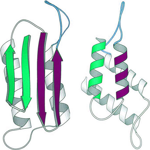
VINOTHAN: Well viruses certainly are interesting in medicine and biology because we want to understand how to prevent disease. But physicists have been studying viruses for a long time and they have a very beautiful structure to them. Viral capsids have a very beautiful structure to them and a very elegant geometrical structure. At the same time, from a physicists point of view, it’s one of the simplest biological systems we can look at. The reason that it’s so simple is that viruses are simple organisms. There’s not a whole lot of processes going on there that are active—not a whole lot of motor proteins, for example, that are moving things around physically. A lot of the classic tools we use in physics we can start to use to analyze viruses such as statistical mechanics and thermodynamics, but yet at the same time, viruses can reproduce themselves, they can inject themselves into a cell, they can disassemble on cue, they can do all sorts of fantastic things that are much more complex than the usual physical systems we look at.
Interviewer: So it’s relatively well understood once things are assembled, but how they assembled is less well understood?
VINOTHAN: Structural biologists can tell us how two proteins are shaped, what every atom on their surface looks like, and they can tell us exactly what happens as they bind together. They can tell us exactly where the binding sites are, what amino acids are involved in the binding, and even—to some extent—when they’re bonded together, how the structures of the two proteins change, after they bond together. But what we don’t understand very well is the process of many of these proteins coming together to form some ordered structure. And that’s what I think is one of the interesting things that we can explore using the tools that we’re looking at in my lab and using new tools that might be developed in molecular and structural biology—looking at the dynamics of how biological systems self-assemble.
Interviewer: Can you some more detail about what a virus is?
VINOTHAN: So a virus exists in this very unique space between what we would consider to be dead and something we would consider to be alive. They can reproduce themselves, clearly, they have genetic material, but at the same time they don’t take in food, they don’t excrete waste, they don’t have any of the properties that we normally think of as properties of a biological living organism. They are a very simple organism at best. Physicists like the fact that they’re not completely dead because things that are completely dead are the things we usually study. Atomic systems that form crystals are completely dead, they do interesting things, but we’d like to take it up a step and start looking at things that are sort of part way alive, and a virus is like that.
Interviewer: So let’s move to the experimental side of your work. What are the experiments that you’re working on?
VINOTHAN: Structural biologists can tell us how two proteins are shaped, what every atom on their surface looks like, and they can tell us exactly what happens as they bind together. They can tell us exactly where the binding sites are, what amino acids are involved in the binding, and even—to some extent—when they’re bonded together, how the structures of the two proteins change, after they bond together. But what we don’t understand very well is the process of many of these proteins coming together to form some ordered structure. And that’s what I think is one of the interesting things that we can explore using the tools that we’re looking at in my lab and using new tools that might be developed in molecular and structural biology—looking at the dynamics of how biological systems self-assemble.
Interviewer: What is the simplest experiment you perform?
VINOTHAN:The first experiment we started with was just looking at identical spherical particles with a very short-range interaction that we made by engineering the fluid around them so that they had this particular interaction. It’s called a depletion interaction. The particles attract each other at a range, which is much, much smaller than their diameter, so essentially they can come together and when they touch they stick. We can adjust the magnitude of that interaction, so the depth of that interaction, how much energy is in that binding. Then we simply made a whole bunch of clusters and micro wells with different particle numbers that were trapped inside micro wells and looked at what structures formed. This system I consider to be very simple.
People have been looking at pairs of particles for a long time to understand the parent reactions, and people had also been looking at bulk systems, large numbers of particles to try to understand how crystals formed for a very long time, and glasses, and other sorts of macroscopic bulk phases. But this, in between range, was a different area, a new area, to study, and we found some interesting results.
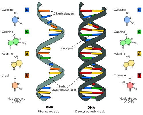
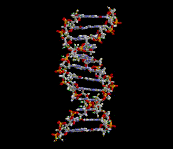
Interviewer: Use of DNA is an area of research that your lab is interested in?
VINOTHANYes. The latest thing we’ve been doing is looking at slightly more complex systems, and the way we’ve made the system more complex is that we’ve gone from using single spheres, that are all identical, to two “species” of spheres. If we call them species A and species B, perhaps A can combine to B but A cannot bind to A, for example. And the question we want to ask is when you have these kinds of binding rules, and certain biological systems do have these kinds of binding rules, what are the kinds of structures that form? We think this is the simplest extension we can go with to hopefully observe some new structures.
Interviewer: This is extending the research into the biological area.
VINOTHAN:The way we engineer these interactions is actually by using biology itself. We cover the surface of the particles with DNA, and DNA is a very well-understood biological molecule, and by using DNA we can engineer two particles that interact with each other, or we can engineer systems where you have two species: A and B, where A can bind to B but A does not bind to A.
DNA is maybe the best understood biological molecule around, and it’s easy to design a sequence of DNA, a strand of DNA, with a particular energy of binding to another strand of DNA. It’s easy to compute that. And in fact you can go on the Internet and order some nucleotide sequence in exactly the sequence you want and have it delivered to you the next day.
Interviewer: There’s no way you can see DNA with the optical regime that you’re using. You’re just interested in its effect on the colloid particles.
VINOTHAN:The DNA is just a way to engineer the kinds of interactions we want. We’re not studying the DNA assembly itself. We’re really seeing what happens to the particles. The DNA is on the order of nanometers in size, whereas the particles we look at are maybe a thousand nanometers in size. When we actually look at our system, all we see are the particles interacting, we don’t see the DNA at all, but we know it’s there because we can label the particles that have DNA on their surface, for example, and see them show up brightly in our fluorescence microscope.
Interviewer: Using Geomags (modeling toys) to explain results of experiments.
VINOTHAN: When we did experiments where we took spherical particles, all the same kind of particle, and stuck them into micro wells and let them stick together, when we had six particles we find that we get two different. At two, three, four and five particles there’s only one structure that forms, so six particles is the first interesting case. One of those structures is an octahedron, it’s a very symmetric shape, it’s called a platonic solid. And the other is basically three tetra-hedron which are stuck base to base, which is much less symmetric than an octahedron. Both have the same number of bonds between the spheres, so these little silver balls are like the centers of my polymer spheres in the experiment, and these things connecting them are where two spheres touch. Both of these have the same number of rods, twelve rods, that are connecting the spheres. In our experiment they actually have the same energy, but what we find is that this one, the less symmetric one, occurs a factor of 24 times as often as this one—much more often. So the question is why? And the answer must not have anything to do with energy because they both have the same energy. And so the only answer that seems to work is that it depends on entropy.
Interviewer: Would you say that Nature usually favors symmetry?
VINOTHAN: It was very surprising to us that this highly symmetric structure would occur so much less frequently than this asymmetric structure. In retrospect it makes perfect sense because we now have models that explain all the contributions to the entropy of these two things, but in many fields of condensed matter physics, what tends to form, for atomic systems for example, is the more symmetric structure. In biological systems as well, for viruses, viruses are highly symmetric. They have very high symmetry. So we were surprised when we found so much of this. And we found that rule applies even at larger numbers of particles as well, the highly symmetric structures are extremely unfavorable.
This is an example of one of the physical rules that we hope to discover that in this system when we have short-range attraction, asymmetry is strongly favored, or symmetry is highly unfavorable. It’s an unexpected rule, but it makes sense when we think about the contributions to the entropy of symmetry.
Interviewer: How did you observe the structures that formed?
VINOTHAN: The experiment we did to look at the structures of the clusters was, involved a lot of sample preparation, making the micro wells and getting the particles to organize in the right way. But the actual experiment to see them was very simple. We take movies of these clusters under the optical microscope as they diffuse around. Brownian motion tends to cause these structures to rotate around, and so what we’ll do is we’ll look at them under the optical microscope, which basically gives a 2D image—you just get what’s at the focal plate of the microscope. So we’ll have to follow these structures around using the focus knob of the microscope to see where they’re going. But then once we have a movie, we’ll go back and look at it on the computer and we’ll see which spheres are touching each other and then gradually build up a 3D representation of that structure.
Interviewer: Is 3D imaging more useful in your experiments?
VINOTHAN: When we look at the clusters under the optical microscope, what we see is a very thin section of the sample, a thin section near the focal plane of the microscope, and the reason we see a thin section is because we’re using a very high power, high aperture objective to look at the system. To get the maximum amount of resolution possible the trade-off is that we can only see a very thin slice of the sample. That means that we have to continually change the focus in order to see the cluster as it diffuses around. It also means that we can’t see the dynamics of how the cluster forms very well because before things have come together, particles may be in different focal planes at once, so we really need to come up with other microscopy techniques in order to see the dynamics of how things are self-assembling into these clusters.
Interviewer: So 3D imaging helps you a lot more for “real time” observation?
VINOTHAN: Now we look at these systems using different kinds of microscopy in order to look at the dynamics of assembly and look at systems that rearrange quite quickly. We use a technique called digital holographic microscopy, which allows us to take a single 2D image from a microscope, and from that back out all of the 3D information—the structures of all the particles disbursed throughout the volume of the well that we’re looking at.
Interviewer: Let’s go back to the data that comes from 3D and the process of getting that data.
VINOTHAN: The data we get from digital holographic microscopy is called a hologram, and it looks quite different from what you would see under the optical microscope. In the optical microscope, if I have a particle, a single spherical particle, it will look like a bright dot, exactly what you would expect it to look like. In the holographic microscope, it looks like a series of concentric rings, and those rings are the interference pattern from light that’s scattered from the particle, the laser that’s scattered from this particle and the interference of that with the light that just goes straight through the sample and doesn’t bother with the particle at all. And that interference pattern tells us something about where the particle is, along the axis of the beam that it’s going through, so it gives us the 3D information about the position of the particle.
Interviewer: You are taking a movie of all these out of focus images, and from that you’ve got a really good 3D image of how the particles are moving within your solution.
VINOTHAN:The advantage of digital holographic microscopy for our systems is that we can back out a full 3D image from a single 2D image, and that means that the technique is fast. So I can take a series of 2D images and very short time scale, say a millisecond or so, and then from that be able to tell what each particle in this entire volume is doing on millisecond timescales, which allows me to avoid the whole problem that I have in optical microscopy where I have to keep changing the focal depth in order to track each of the particles in the system.
So generally we’ll take a few thousands of images spread out over seconds, sometimes over minutes, sometimes longer, and that will give us a full complete picture of how every single particle in the system is moving in all three dimensions as a function of time.
So it’s basically a three dimensional movie of the system.
Interviewer: What are your other tools for recording data?
VINOTHAN: There are some systems that we can’t see under the optical microscope such as proteins. We can’t see what proteins are doing in real time in a fluid under a microscope. They’re just too small to resolve. So we’re very interested in looking at the dynamics of how they assemble. If I am willing to say I can’t get any information about the structure of a protein complex as it assembles, but I want to understand something about the dynamics, then I can still use light to understand something about that. So we use a tool called light scattering. It’s a very simple experiment conceptually. I have a sample full of proteins and I shine a laser on it. In this case I use a highly collimated laser, which means it’s all coming out in a beam that is not diverging or converging at all. I simply look at the amount of light that comes from the sample that is scattered. If a laser is going through the sample I look from an angle to see how much light is coming off of it. The amount of light that’s scattered depends on how big the structures are in solution, and also the amount of light you get as a function of the angle that I’m looking at depends on how big the structures are in solution. So we can get some clues by looking at how this intensity changes over time as to how things are growing inside the sample.
Interviewer: What do these data look like?
VINOTHAN: We have two kinds of light scattering that we do. One is called dynamic light scattering, and the other is called static light scattering. In dynamic light scattering, we measure what’s called a correlation function, and the correlation function says: if I have the intensity at a time here, how related is the intensity at a later time? It’s just a statistical quantity that correlates those two things. And the more things are moving around, the less correlated the intensity will be at a time later than the initial time that I pick. So that tells me something about how fast on average things are diffusing around. The other kind of measurement we do is called static light scattering, and in static light scattering what we measure is intensity as a function of angle. And then what we do is repeat that experiment again and again and again, and what we can see is how that intensity as a function of each angle is changing as a function of time. If we have proteins that are dimerizing together, at the beginning we might see very little intensity at each angle, and as they begin to dimerize together, we’ll see a gradual increase in the intensity. From that we can tell how fast the proteins are coming together, and I can tell something about their interaction strength.
Interviewer: Where do you think your research will go, especially related to viruses?
VINOTHAN: Nowadays there are several groups that are working on treatments for viruses that have to do with inhibiting the formation of the capsid, preventing it from forming in the first place, preventing the self-assembly process, so this is a potential direction that in 20, 30 years this research can eventually lead to. If we understand the process of how these things self-assemble then we might have some ideas about how to disrupt that process and keep the virus from propagating and from replicating itself and infecting.
Coming from the perspective of nanotechnology, where we spend a lot of time trying to build things through self-assembly, that that should be relatively straightforward because we find that it’s actually very easy to disrupt the self-assembly process, but you have to know the right way to do it. We have to understand something about that process in order to disrupt it, so I think that having some idea of what’s going on with the dynamics will really help enable these new techniques for inhibiting capsid formation.
Interviewer: What does the future hold for your research?
VINOTHAN: So I think now we’re just starting to understand some of the new physics that surrounds the idea of what I would call complexity. When you have anything more than two particles in a system we see that it forms all sorts of interesting shapes and we see all sorts of interesting behavior and this phenomenon of self-assembly, understanding it, harnessing it for nano technology, and at the same time understanding it in order to do something useful in medicine, relies on understanding complexity, this physical problem of what happens when you have many particles coming together at once.
I think that these colloidal systems are really nice because you can tune things so that you can exactly control the interactions and can put them in a variety of different situations to just see what happens. What we’ve seen so far is that interesting things happen, interesting things that we wouldn’t necessarily have predicted before. In the future I think we’ll be able to use some of these physical principles that we gathered from looking at these model systems in order to do make interesting materials in nano technology, and at the same time maybe to actually think about diseases in biology as well, at least those pertaining to viruses.
For now, at least in the near term, and in fact in the long term too, I think there’s value to doing these very simple experiments where we just explore and see what happens, experiments based on discovering new phenomenon. And I think that having all of these paths that are a very applied path and at the same time they’re very exploratory paths is vital for making sure we succeed.